INTRODUCTION
Crohn’s disease (CD) is a chronic inflammatory disease characterized by ulcers of the small and large bowels, progressing to the development of complications including strictures and fistulae. The treat-to-target approach is the treatment strategy that is currently recommended in treating inflammatory bowel disease (IBD) patients, where not only clinical remission but also endoscopic remission is set as a formal target [1]. Endoscopic remission, in this context, is defined as that which is confirmed by ileocolonoscopy, the preferred method worldwide for assessing mucosal healing in patients with CD. However, patients can often manifest with more proximal lesions that cannot be reached by ileocolonoscopy. We have previously shown that evaluation of small bowel mucosal healing more proximal to the terminal ileum using balloon-assisted enteroscopy (BAE) is important, as active (ulcerative) lesions in this area are associated with a higher rate of clinical relapse, hospitalization, and surgery [2]. Nonetheless, BAE is an invasive procedure with potential risks, high cost, and limited accessibility, as are other available imaging techniques, such as capsule endoscopy, magnetic resonance enteroscopy (MRE), and bowel ultrasound.
Compared to these imaging modalities, biomarkers are noninvasive and less expensive, and can be performed in non-specialized centers. C-reactive protein (CRP) and fecal calprotectin are 2 such biomarkers that have been well established for use in IBD patients. Recently, the CALM trial has shown that tighter disease control based on these biomarkers irrespective of symptoms led to improved prognosis and endoscopic remission [3]. They have been defined as formal targets in the aforementioned treat-to-target approach. However, studies focusing on the use of fecal calprotectin with regards to small bowel activity have been limited, with mixed results, suggesting room for improvement [4-7]. Fecal hemoglobin has also been reported to be useful in CD, with some reports suggesting similar accuracy as fecal calprotectin [8], while others reporting a lower specificity in terms of small bowel activity [9]. Leucine-rich alpha-2 glycoprotein (LRG) is a novel biomarker that has recently been approved for use in the diagnosis and monitoring of IBD patients [10]. Unlike CRP which is induced mainly by interleukin (IL)-6, LRG expression is also induced by tumor necrosis factor-α, IL-1β, and IL-22, enabling a more sensitive reflection of a wider range of inflammation. It has been reported to be a sensitive marker for detecting small intestinal activity and transmural healing in CD patients [11]. Our previous study involving CD patients undergoing BAE showed that LRG was significantly more accurate in detecting endoscopic activity compared to CRP [12]. Several other studies have also shown accuracy of LRG in assessing for small bowel lesions using BAE [13], and capsule endoscopy [14]. Shinzaki et al. [15] showed that LRG correlates better with endoscopic activity than fecal calprotectin or CRP, in CD patients evaluated using colonoscopy. However, no previous study has directly compared these existing biomarkers in terms of monitoring for endoscopic activity including the small bowel.
Several studies have combined biomarkers for IBD in the past. Combining fecal calprotectin, CRP, and clinical activity scores have been reported to be useful for monitoring endoscopic activity in both CD and ulcerative colitis [16,17], however, no reports have evaluated their combined use for detecting endoscopic activity in CD evaluated by enteroscopy.
We investigated which of the existing biomarkers best reflects endoscopic activity in CD patients evaluated using BAE, and whether combining these biomarkers could improve their accuracy.
METHODS
1. IRB Approval
The study was conducted in accordance with the Declaration of Helsinki. The study was approved by the Ethics Committee of Tokyo Medical and Dental University (IRB No. M2019-11). Written informed consent was obtained from all patients.
2. Study Population
We performed BAE (SIF-H190; Olympus Medical Systems, Tokyo, Japan) in 104 consecutive patients with ileal and ileocolonic type CD from October 2021 to August 2022 at our hospital (Tokyo Medical and Dental University, Japan), a tertiary referral hospital, for assessment of known or suspected small bowel lesions. Clinical and endoscopic data were prospectively collected. MRE results were also collected if performed within 8 weeks of undergoing BAE (n = 32). Hemoglobin, platelet count, CRP, and LRG measurements were made together with other routine blood at outpatient clinic within 8 weeks of performing BAE, before any changes were made to treatment from time of endoscopy. Both CRP and LRG were measured using commercially available latex agglutination kits (N-Assay LA CRP-T kit, Nittobo Medical, Tokyo, Japan; Nanopia LRG, Sekisui Medical, Tokyo, Japan) within the hospital. Patients were asked to bring a fecal sample on the day of BAE, which was collected at home before initiating bowel preparation. Fecal samples were analyzed for hemoglobin and calprotectin within the hospital (EIKEN OC-sensor; Eiken Chemical Co., Ltd., Tokyo, Japan). All patients filled the diagnostic criteria outlined by the Japanese Ministry of Health, Labour and Welfare [18]. Exclusion criteria included patients with colostomy/ileostomy, those with abdominal abscesses, those who were intolerant of or contraindicated to BAE, those on nonsteroidal anti-inflammatories, and those with confirmed intestinal infections or malignancies. Patients were prospectively followed for at least 6 months for evaluation of clinical outcome (hospitalization, surgery, and relapse related to CD). Relapse was defined as clinical or endoscopic activity which required change of treatment by the physician in charge.
3. Imaging Modalities
The protocol for BAE at our institution has been described previously [19]. Retrograde insertion was performed for all cases. Scope insertion was terminated when large ulcers were detected in order to avoid complications. Endoscopic evaluations were performed using the modified Simple Endoscopic Score for CD (modified SES-CD) as described previously [20]. In short, this excluded “stenosis” from the original SES-CD which includes 4 endoscopic variables (size of ulcers, proportion of ulcerated surface, proportion of affected surface, and stenosis), as we aimed to focus on active disease. The small bowel was divided into 3 segments; terminal ileum, proximal ileum, and jejunum. Each variable was scored 0 to 3 for each segment, and the modified SES-CD was calculated as the sum. Anastomotic ulcers were also included. Endoscopists blinded to the results of the laboratory tests evaluated the score.
We performed and evaluated MRE as outlined previously [11]. A 3.0-T scanner (FUJIFILM Healthcare Corporation [formerly HITACHI], Tokyo, Japan) was used with oral and intravenous contrast. All imaging procedures covered the entire small bowel. A radiologist, who was blinded to the laboratory test results, determined the simplified magnetic resonance index of activity [21]. Transmural activity was defined as a maximum segmental simplified magnetic resonance index of activity ≥ 4, in line with our previous study [11].
4. Study Design
Primary outcome was set as the relationship between each biomarker and the presence of intestinal ulcers identified at BAE. “Presence of ulcer” was defined as presence of a mucosal defect ≥ 0.5 cm, as defined in SES-CD. The effect of combining biomarkers was also evaluated. Subanalyses were performed for patients with absence of colonic ulcers, extraintestinal manifestations, active anal disease, and patients who also underwent MRE.
5. Statistical Analysis
Between-group differences in continuous variables were assessed by the Mann-Whitney test. The Fisher exact test was used to analyze between-group differences for categorical variables using 2 × 2 contingency tables. The relationship between the presence of ulcers and each biomarker was examined using receiver operating characteristic (ROC) curves and the area under the curve (AUC). The optimal threshold value for each biomarker was calculated using the Youden index. Logistic regression analysis was used to determine the relationship between different biomarkers and the presence of ulcers. The presence of ulcers was the outcome variable (or dependent variable) (i.e., a binomial variable taking the value 1 if presence of ulcer ≥ 0.5 cm and the value 0 if < 0.5 cm). Each biomarker with their calculated optimal cutoffs (CRP, LRG, hemoglobin, platelet, fecal hemoglobin, and fecal calprotectin) were employed as explanatory variables (or independent variables). Univariable analysis was used to identify candidate predictors. Then, a multivariable model was fitted. All variables with P<0.05 were retained in the model. Confidence intervals were calculated using the profile likelihood method. The Spearman correlation coefficient was used to analyze the strength of correlation between various biomarkers and modified SES-CD. To evaluate patient prognosis, the Kaplan-Meier method and the log-rank test were used to calculate cumulative end point-free rates, whereas Cox proportional hazards model was used to evaluate the risks for prognosis. The level of statistical significance was set at a P-value of < 0.05. Box plot figures present median (line), interquartile range (box), range (whiskers), outliers (open circles), and extreme outliers (stars). GraphPad Prism 9.5.1 (GraphPad Software, San Diego, CA, USA) and R version 3.3.0 (R Foundation for Statistical Computing, Vienna, Austria) were used.
RESULTS
1. Patient Characteristics and Endoscopic Findings
The main characteristics of the 104 patients included in the study are shown in Table 1. Their disease classed by the Montreal classification included 26 patients with inflammatory disease (B1), 55 patients with stricturing disease (B2), and 23 patients with penetrating disease (B3). The majority of the patients (87%) were treated with biologics including infliximab, adalimumab, ustekinumab, and vedolizumab. Most of the patients were in clinical remission, with a median Crohn’s Disease Activity Index of 57. The small bowel was intubated in all 104 patients, and the proximal ileum was reached in almost all of the cases (96%). Median length of small bowel visualized from the ileocecal valve (or anastomotic site in the case of postsurgical patients) was 50 cm. The extent of intubation was often limited by the presence of a stricture or difficulty in manipulation. There were 55 patients in whom ulcers were identified at BAE; of whom 52 patients had small bowel ulcers, with 19 patients having both small bowel and colonic ulcers.
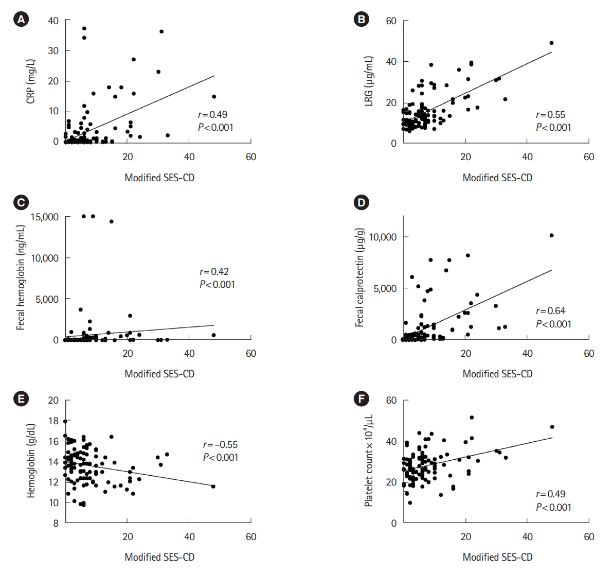
2. Biomarkers Detect and Correlate with Endoscopic Activity in CD Patients
There was a significant difference in all blood and fecal biomarker values between CD patients in whom ulcers were observed by BAE compared to those without (Fig. 1). CRP, LRG, fecal hemoglobin, fecal calprotectin, and platelet count were significantly increased in those with ulcers, whereas hemoglobin was significantly decreased in those with ulcers. We then evaluated the correlation of endoscopic activity detected at BAE with each of the biomarkers. All biomarkers significantly correlated with modified SES-CD, a score for endoscopic activity which evaluates the small bowel in detail as well as the colon (Fig. 2). Out of the 6 biomarkers evaluated, fecal calprotectin showed the strongest correlation with endoscopic activity (r=0.64, P<0.001), while CRP, LRG, fecal hemoglobin, hemoglobin, and platelet count all showed moderate correlation (r=0.49, r=0.55, r=0.42, r=-0.55, r=0.49 respectively, all P<0.001).
3. Fecal Calprotectin and LRG Detect Endoscopic and Transmural Activity
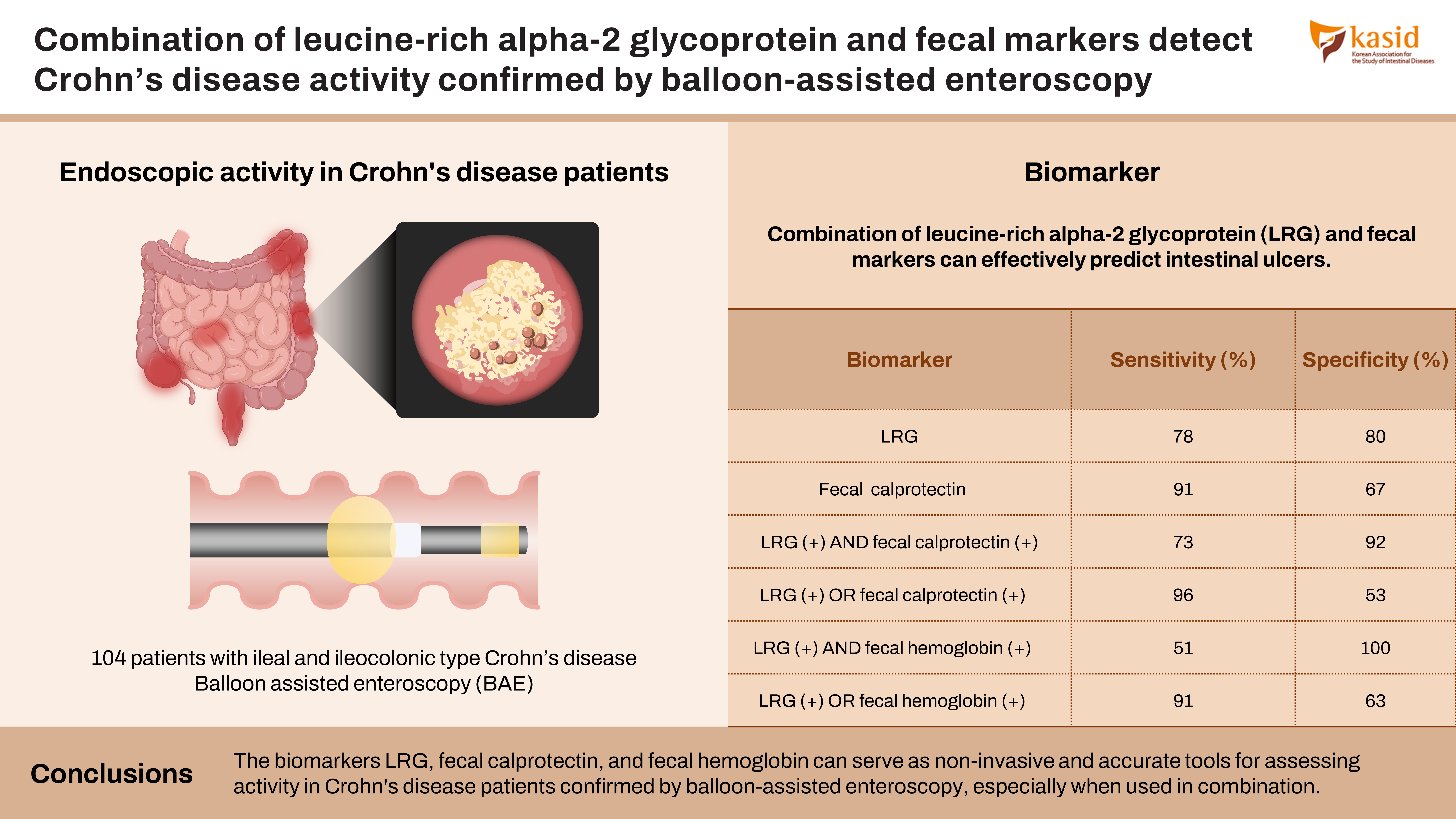
We evaluated the diagnostic ability of the biomarkers in detecting the presence of intestinal ulcers detected on BAE, by analyzing the AUC of ROC curves (Table 2). The AUCs for fecal calprotectin and LRG were 0.853 and 0.841 respectively, and were higher than the AUCs for CRP, hemoglobin, platelet count, or fecal hemoglobin (0.813, 0.653, 0.638, 0.761, respectively). The optimal threshold value for each marker was calculated from the respective ROC curves using the Youden index. Fecal calprotectin showed a high sensitivity of 91%, specificity of 67%, positive predictive value (PPV) of 76%, and negative predictive value (NPV) of 87% at a cutoff value of 151 μg/g. LRG also showed a high sensitivity of 78%, specificity of 80%, PPV of 80%, and NPV of 76% at a cutoff value of 13 μg/mL. Because our ulcer population consisted of patients with both small bowel and colonic ulcers, in addition to patients with active extraintestinal manifestations and active anal disease, we performed subanalysis excluding 22 patients with colonic ulcers, 13 patients with active anal disease, and 2 patients with active extraintestinal manifestations (n = 75). LRG and fecal calprotectin still showed high accuracy for detecting solely small bowel ulcers compared to those without, with an AUC of 0.805 and 0.784 respectively, with CRP also yielding a comparable result with an AUC of 0.785 (Table 3).
Because BAE cannot assess the whole of the small bowel, nor detect for transmural inflammation, subanalysis was performed for the 32 cases that also underwent MRE as well as BAE (Supplementary Table 1). LRG and fecal calprotectin were accurate as markers to detect cases where activity was seen on either BAE or MRE, compared to those without any activity on either modality, with an AUC of 0.836 and 0.806 respectively. Although not statistically significant, biomarker levels tended to be lower in cases of endoscopic remission where MRE also confirmed remission (radiological remission), compared to cases in endoscopic remission where MRE showed activity (Supplementary Fig. 1).
4. LRG and Fecal Biomarkers Independently Predict Ulcers
In order to determine the relationship between the biomarkers, we analyzed the correlation between each of the biomarkers (Supplementary Table 2). LRG showed the strongest correlation with CRP (r=0.78, P<0.001), another blood biomarker, whereas fecal calprotectin showed the strongest correlation with fecal hemoglobin (r=0.64, P<0.001), a fecal biomarker. There was a tendency for the correlation to be weaker between blood and fecal biomarkers. Although LRG and fecal calprotectin were both accurate in detecting ulcers, these results suggested that blood and fecal biomarkers may detect slightly different inflammatory profiles. We therefore determined the relationship between each of the biomarkers with ulcers found on BAE using logistic regression. Univariable analysis showed statistical significance for all biomarkers, however only LRG ≥ 13 μg/mL (odds ratio, 4.946; P=0.033), fecal hemoglobin ≥ 41 ng/mL (OR, 5.824; P=0.016), and fecal calprotectin ≥ 151 μg/g (odds ratio, 6.082; P=0.012), were independent predictors for the presence of ulcers on BAE (Table 4).
5. Combination of LRG and Fecal Biomarkers Accurately Detect Ulcers on BAE
We therefore hypothesized that combining the different types of biomarkers could improve their accuracy. The ability to detect ulcers on BAE using various combinations of LRG, fecal hemoglobin, and fecal calprotectin, with their pre-determined optimal cutoff values, were determined (Table 2). As predicted, combining LRG, a serum biomarker, together with each of the fecal biomarkers improved the accuracy. Dual-positivity for LRG and fecal calprotectin predicted the presence of ulcers with an improved specificity of 92% and PPV of 91%, whereas having either a positive LRG or fecal calprotectin showed an improved sensitivity of 96% and NPV of 93%. The combination of LRG and fecal hemoglobin showed a similar improvement in accuracy; dual-positivity for LRG and fecal hemoglobin predicted ulcers with an improved specificity of 100% and PPV of 100%, whereas having a positive LRG or fecal hemoglobin showed an improved sensitivity of 91% and NPV of 86%. There was no marked added improvement when all 3 of the biomarkers were combined.
6. Combination of LRG and Fecal Biomarkers Predict Patient Prognosis
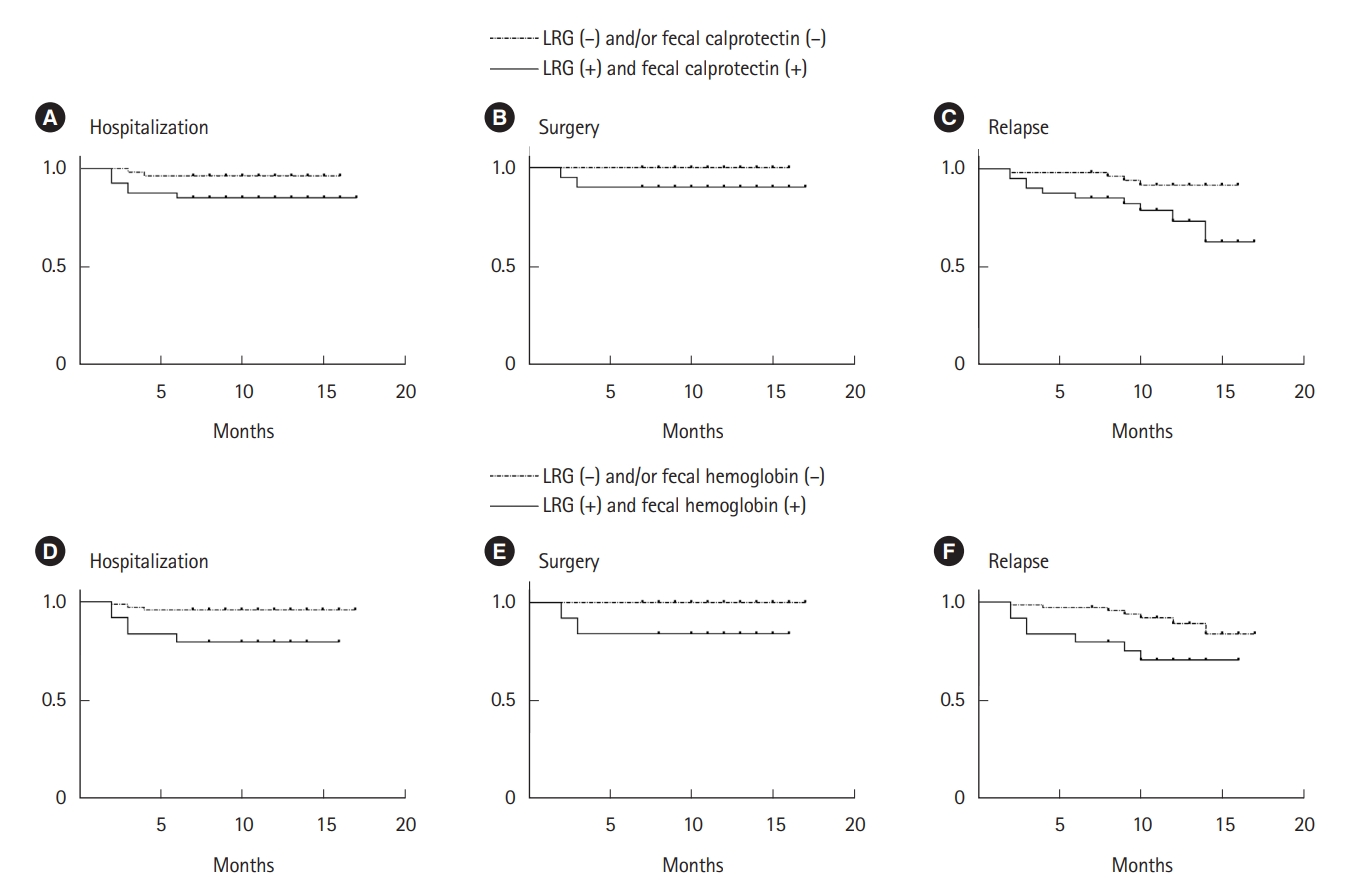
Finally, we sought to determine the association of biomarkers with patient prognosis. Out of the 104 patients followed after a median of 11 months (range, 0-17 months), 8 patients were hospitalized, 4 patients underwent surgery, and 14 patients relapsed. Relapse was defined as those requiring a change of treatment. Kaplan-Meier analysis showed that patients with dual-positivity for LRG and fecal calprotectin demonstrated a poorer outcome-free rate in terms of hospitalization, surgery, and relapse, compared to those with either low LRG and/or low fecal calprotectin (Fig. 3A-C). Cox proportional hazard regression showed a significant hazard ratio of 3.846 (95% confidence interval, 1.285-14.03; P<0.05) for relapse in the dualpositive group. Similar results were obtained with LRG and fecal hemoglobin, where those who were dual-positive demonstrated a poorer outcome-free rate compared to those with either low LRG and/or low fecal hemoglobin (Fig. 3D-F). Cox proportional hazard regression showed a significant hazard ratio of 3.325 (95% confidence interval, 1.134-9.755, P<0.05) for relapse in the dual-positive group.
DISCUSSION
Biomarkers are recommended for monitoring CD activity; however, their accuracy has been debated, especially in detecting small bowel activity [4,9]. This is the first study comparing the major existing biomarkers for CD in terms of monitoring for endoscopic activity evaluated using BAE, as well as the added value of their use in combination. We showed that LRG and fecal biomarkers, especially fecal calprotectin, are both accurate biomarkers on their own in detecting and correlating with endoscopic activity, but their accuracy is further improved when used in combination. Dual positivity for LRG and either fecal biomarker shows high specificity and PPV, suggesting that when a patient tests positive for both markers, there is a high likelihood that the patient will have endoscopic activity detected with BAE. Conversely, the sensitivity and NPV are high if LRG or either fecal biomarker is positive, indicating that if a patient tests negative for both of these markers, there is a low likelihood of the patient having endoscopic activity detected with BAE. We also showed that the combined use of LRG and fecal calprotectin or fecal hemoglobin predicts the clinical outcome of CD patients better than each on its own. Decreased hemoglobin and increased platelet count are known as robust inflammatory markers in IBD and were included in the study, however, their abilities to detect endoscopic activity were poor compared to the other markers. Although CRP showed a relatively high AUC, it did not emerge as an independent predictor as a result of multivariate analysis.
In this study, we included patients with both ileal and ileocolonic type CD, and set the primary outcome to include any intestinal ulcers confirmed on enteroscopy, assessing for both the small bowel and the colon. We also decided to include patients with active anal disease and extraintestinal manifestations, to reflect the patient population encountered in actual clinical practice. As reported previously [22], small bowel healing is much more difficult to achieve compared to colonic healing in cases of ileocolonic CD; rarely resulting in solely colonic residual ulcers. This is confirmed in our study where out of the 64 patients with ileocolonic type CD, only 3 patients had residual colonic ulcers alone, compared to 15 patients with ulcers only in the small intestine, and 19 patients with ulcers in both the small bowel and colon. In clinical practice, we do not only deal with ileal type CD but also ileocolonic CD, with varying degrees of small bowel and colonic ulcers, in addition to patients with perianal lesions and extraintestinal manifestations. We, therefore, included these patients in our main analysis as any biomarker will have to perform well even in patients with these commonly seen complications. We hypothesized that including these “extra” inflammatory burdens would result in a lower specificity for the biomarkers, especially the serum markers, however on the contrary, our subanalysis excluding these sets of patients to assess for purely small bowel ulcers resulted in a lower accuracy for detecting endoscopic activity (Table 3). This may be because of the high rate of endoscopically active lesions in this group of patients (24 out of 29 patients had endoscopically confirmed ulcers). We were able to show that these biomarkers will be useful in patients in a real-life clinic setting.
LRG is a serum biomarker which is induced by systemic inflammation, providing an advantage as it can reflect transmural inflammation in CD, as previously reported [11]. However, this feature is also a disadvantage as it responds to non-specific inflammation other than IBD [23]. On the other hand, fecal biomarkers are more specific towards bowel mucosal inflammation. Our subanalysis of a limited number of patients who also underwent MRE showed that CRP and LRG were better markers for detecting transmural activity than fecal hemoglobin or fecal calprotectin. This difference in mechanism between the markers is possibly the reason for their improved accuracy in detecting endoscopic activity when used in combination, complementing their weaknesses. Whilst our study could not identify particular parameters associated with false positivity/negativity of individual biomarkers, future larger-scale studies are warranted to address this question.
Although fecal hemoglobin was less accurate than fecal calprotectin in detecting endoscopic activity compared to fecal calprotectin (AUC: 0.761 vs. 0.853, P=0.05), the accuracy of fecal hemoglobin improved when combined with LRG. In clinical settings where fecal calprotectin measurement is not available, or when LRG and fecal calprotectin cannot be simultaneously measured due to insurance restrictions, as is the case in Japan, the combined use of LRG and fecal hemoglobin can be an alternative monitoring method.
We also showed for the first time that combining LRG and fecal biomarkers can better predict the clinical outcome of CD patients in the long term. Patients who are dual-positive for LRG and fecal calprotectin or hemoglobin should be considered for endoscopic evaluation including the small bowel and assessed for change in treatment, as they are at higher risk for hospitalization, relapse, and surgery.
The limitation of our study is that it was conducted at a single tertiary referral hospital in Tokyo. The length of small intestine intubated varied in each individual, with possibility that the modified SES-CD scores do not accurately reflect the endoscopic activity of the whole intestine. Our study also did not consider the areas not intubated by enteroscopy (i.e., the proximal small intestine, stomach, and duodenum). We, therefore, performed subanalysis on patients who also underwent MRE, however, the number of cases was limited. The patient population consisted of mostly biologic-treated patients in clinical remission, therefore may not be representative of the whole patient population. Blood and fecal biomarkers were not necessarily taken on the same day as each other, and not simultaneously with BAE or MRE, though tests were all conducted in a limited timespan before change of treatment. Because patients enrolled were mostly in remission with stable disease undergoing routine elective endoscopy, we predicted that disease activity would not change markedly within the 8-week time period.
In conclusion, the combined use of LRG and fecal biomarkers detects with high accuracy the presence of endoscopic activity evaluated by BAE in CD patients. The use of these biomarkers in clinical practice may enable earlier intervention, leading to better prognosis.