Lymph node macrophages: drug-related reaction or infectious-lesion?
Article information
The involvement of myeloid-derived cells in IBD pathogenesis both in gut inflammatory loci and lymphoid organs is well known, as reviewed by Kim et al. [1] We had recently the opportunity to identify groups of macrophages in a lymph node detected on a cholecystectomy specimen. The patient (man, 47 years) was admitted for abdominal pain, hyperleukocytosis and elevated CRP. The medical history revealed CD (diagnosed at the age of 33 years) with recurrent anal fissures (treated by 5 surgeries). The medical treatment comprised initially mesalazine and more recently ifliximab and azathioprine, azathioprine being associated with abdominal pain. The CT-scan at the time of admission showed a lithiasic gallbladder with thickened wall. A coelioscopic cholecystectomy was performed. The resected specimen consisting in 2 fragments measuring 7.4 and 4.4 cm. The microscopy diagnosis was that of subacute cholecystitis with foci of intestinal metaplasia. The lymph node detected on the surgical specimen, contained several extravascular macrophages, PAS and CD68 positive (Fig. 1). The immunohistochemistry for the Tropheryma whipplei was negative.
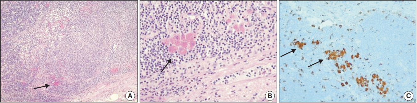
The gallbladder lymph node contained several extravascular PAS- (A, B, H&E) and CD68-positive macrophages (C, immunohistochemical staining) (arrows). Original magnification ×2.5 (A), ×40 (B), and ×20 (C).
Extravascular macrophage accumulations in lymph nodes are rare [2]. Transient accumulations are reported in rhesus monkey models, during acute SIVmac239 virus infection [3]. In the case we report, the histogenesis of the lymph node macrophage accumulation is difficult to precise. An infectious origin was ruled out as based on the medical history and negativity of T. whipplei immunohistochemistry. Remained the hypothesis of drug-related lesions, both ifliximab and azathioprine being reported to influence the macrophage number. While the impact of ifliximab in CD is varied, reported to result both in CD68-positive macrophage reduction [4] and in regulatory macrophage induction [5], including with occurrence of a macrophage activating syndrome [6], azathioprine is reported to affect the number of peritoneal macrophages in mice models, when administered at high dose and over a long period [7]. However, to note would also be the possibility of a pre-granulomatous, macrophage reaction, as related to influx of IBD myeloid-derived cells [1], associated or not to the potential impact of the medical treatment.
In conclusion, macrophage accumulations may occur in the gallbladder lymph node in the context of IBD, possibly as related to medical treatments with infliximab and/or azathioprine.
Notes
FINANCIAL SUPPORT
The authors received no financial support for the research, authorship, and/or publication of this article.
CONFLICT OF INTEREST
No potential conflict of interest relevant to this article was reported.
AUTHOR CONTRIBUTION
Writing and approval of final manuscript: Adriana HandraLuca.