Intest Res Search

CLOSE


Intest Res > Volume 21(1); 2023 > Article
Oh, Hong, Ham, Lee, Park, Yang, Yoon, Kim, Choi, Ye, and on behalf of the Korean Association for the Study of Intestinal Diseases: Real-world effectiveness and safety of ustekinumab induction therapy for Korean patients with Crohn’s disease: a KASID prospective multicenter study

Abstract

Background/Aims

We investigated the real-world effectiveness and safety of ustekinumab (UST) as induction treatment for Koreans with Crohn’s disease (CD).

Methods

CD patients who started UST were prospectively enrolled from 4 hospitals in Korea. All enrolled patients received intravenous UST infusion at week 0 and subcutaneous UST injection at week 8. Clinical outcomes were assessed using Crohn’s Disease Activity Index (CDAI) scores at weeks 8 and 20 among patients with active disease (CDAI ≥150) at baseline. Clinical remission was defined as a CDAI <150, and clinical response was defined as a reduction in CDAI ≥70 points from baseline. Safety and factors associated with clinical remission at week 20 were also analyzed.

Results

Sixty-five patients were enrolled between January 2019 and December 2020. Among 49 patients with active disease at baseline (CDAI ≥150), clinical remission and clinical response at week 8 were achieved in 26 (53.1%) and 30 (61.2%) patients, respectively. At week 20, 27 (55.1%) and 35 (71.4%) patients achieved clinical remission and clinical response, respectively. Twenty-seven patients (41.5%) experienced adverse events, with serious adverse events in 3 patients (4.6%). One patient (1.5%) stopped UST therapy due to poor response. Underweight (body mass index <18.5 kg/m2) (odds ratio [OR], 0.085; 95% confidence interval [CI], 0.014-0.498; P=0.006) and elevated C-reactive protein at baseline (OR, 0.133; 95% CI, 0.022-0.823; P=0.030) were inversely associated with clinical remission at week 20.

Conclusions

UST was effective and well-tolerated as induction therapy for Korean patients with CD.

INTRODUCTION

Although medical treatment of Crohn’s disease (CD) has significantly improved after the introduction of anti-tumor necrosis factor (TNF) agents, approximately one-fifth of patients do not initially respond to anti-TNF agents (primary nonresponse), and 23% to 46% of patients eventually lose their responses to anti-TNF agents (secondary loss of response) [1,2]. Moreover, the response rate to second- or third-line anti-TNFs has been reported to be lower among anti-TNF-failed patients with inflammatory bowel disease (IBD) than among biologic-naïve patients, and a lack of response necessitates switching to drugs with mechanisms of action different from those of anti-TNFs [3,4].
Ustekinumab (UST) is a fully human immunoglobin G1 monoclonal antibody that blocks the p40 subunit of interleukin (IL)-12 and IL-23 [5]. UST was first approved as a treatment for psoriasis [6]. IL-12 and IL-23, which modulate T-helper (Th) 1 and Th 17 cells, are key proinflammatory cytokines in the pathogenesis of CD [7,8]. Therefore, UST was expected to be efficacious in the treatment of CD by blocking the inflammatory cascade mediated by IL-12 and IL-23. UST has been proven to be efficacious as induction and maintenance therapy for patients with moderate to severe CD in randomized controlled trials [9-11]. UST induction therapy (6 mg/kg) has been shown to yield clinical responses in 38% to 57% of patients and clinical remission in 21% to 40% of patients with CD at week 8 [11]. Additionally, UST induction therapy has been shown to significantly reduce C-reactive protein (CRP) and fecal calprotectin levels [11]. UST has since been associated with clinical benefits as an induction therapy for patients with CD in several multicenter studies from Europe and North America [12-21]. In addition to the clinical response, it has also been shown to be effective for inducing short-term biomarker and/or endoscopic responses [12,14,15,17,18,21]. In a pooled analysis of phase 2 and 3 placebo-controlled trials for patients with IBD, the UST treatment group did not show any differences in terms of development of adverse events (AEs), serious AEs (SAEs), infections, serious infections, and malignancies excluding nonmelanoma skin cancer compared with the placebo group through 1 year [22].
However, most studies on UST treatment for CD have been performed in the Western world; therefore, real-world data on the clinical benefits of UST for Asian patients with CD are still lacking [23-25]. As the incidence and prevalence of IBD in Asian countries have been increasing during the last few decades [26-29], different genetic and phenotypic characteristics have been reported among Asian IBD patients compared with Western IBD patients [30-32]. Unlike with Caucasian patients, there have been conflicting reports on the association between genetic polymorphism of IL-23 receptors (IL23Rs) and CD susceptibility among East Asian patients [33,34]. Therefore, the real-world effectiveness and safety of UST among Asian patients must be evaluated and compared with Western data. We investigated the clinical effectiveness and safety profile of UST induction therapy for Korean patients with CD.

METHODS

This was a prospective, observational, multicenter study. Patients with CD who received UST induction therapy between January 2019 and December 2020 at any of 4 participating centers in Korea were prospectively enrolled in the Korean Association for the Study of Intestinal Diseases UST Registry. At week 0, UST was intravenously infused (260 mg for patients with body weight ≤ 55 kg, 390 mg for patients with body weight > 55 kg but ≤ 85 kg, and 520 mg for patients with body weight > 85 kg), followed by subcutaneous injection of UST 90 mg at week 8. Concomitant therapy with 5-acetylsalicylic acids, immunomodulators, or corticosteroids was permitted. The study protocol was approved by the institutional review board of each participating center, including Asan Medical Center (IRB No. 2018-1388). All study participants provided written informed consent prior to study enrollment.

1. Data Collection

We collected demographic characteristics and CD disease characteristics: age, sex, duration of disease, family history of IBD in first-degree relatives, smoking status, body mass index (BMI), history of bowel resection, previous surgical history for perianal abscess/fistula, concomitant medications, prior use of biologics, Montreal disease location, and Montreal disease behavior [35]. We also collected Crohn’s Disease Activity Index (CDAI) scores and laboratory data, including CRP, serum albumin, and fecal calprotectin levels at baseline (week 0), week 8, and week 20.

2. Outcomes

Clinical outcomes were assessed using CDAI scores at weeks 8 and 20 among patients with active disease (CDAI score ≥ 150) at baseline. Clinical outcomes included the achievement of clinical remission or clinical response. The co-primary outcomes were clinical remission after UST induction therapy (CDAI score < 150 at week 20) or clinical response (reduction in CDAI score ≥ 70 points from baseline [CDAI-70]) at week 20.
Secondary outcomes included corticosteroid-free clinical remission and response, which were defined as clinical remission or response without the need for concomitant systemic corticosteroids (budesonide, prednisone, prednisolone, or methylprednisolone). Changes in biochemical markers, such as serum albumin, serum CRP, and fecal calprotectin levels, were also analyzed. All patients were asked about AEs, including infusion reactions, injection site reactions, exacerbation of CD, infections, hospitalizations, any surgery related to CD, and any potential AEs at regular or unexpected visits. The AEs that resulted in CD-related hospitalization, CD-related bowel surgery, or death were considered SAEs. CD exacerbation was defined as an increase in CDAI ≥ 100 points from baseline, an addition or dose escalation of CD-related drugs, CD-related bowel surgery, or CD-related hospitalization.

3. Statistical Analysis

Patients who received at least 1 dose of UST were included in the effectiveness and safety analyses. Therefore, those who stopped UST or who underwent CD-related bowel surgery before each clinical evaluation were considered to have not achieved clinical effectiveness outcomes. Categorical variables and continuous variables are expressed as numbers with percentages and medians with interquartile ranges (IQRs), respectively. Linear mixed modeling was used to evaluate continuous measurements of CDAI scores and laboratory values, including serum CRP, serum albumin, and fecal calprotectin at weeks 0, 8, and 20. Factors associated with clinical remission and clinical response at week 20 were identified using univariable and multivariable logistic regression analysis. Multivariable analysis was performed using the backward elimination method, including variables with P<0.1 in the univariable analyses. Statistical analyses were performed using SPSS Statistics for Windows, version 21.0 (IBM Corp., Armonk, NY, USA).

RESULTS

1. Patient Characteristics

A total of 65 patients who received UST induction therapy were enrolled. The baseline characteristics of the study participants are summarized in Table 1. Forty-eight patients (73.8%) were men, and the median age at the commencement of UST was 33 years (IQR, 27-45 years). The median disease duration from diagnosis to the first administration of UST was 114 months (IQR, 85-177 months). Fifty-one patients (78.5%) had stricturing or penetrating behavior. Thirty-two patients (49.2%) had perianal disease modifiers, and there were 36 patients (55.4%) each with a history of bowel resection and a surgical history for perianal abscess/fistula. There were 40 patients (61.5%) who were taking concomitant immunomodulators and 5 patients (7.7%) taking concomitant corticosteroids with UST, respectively. Only 7 patients (10.8%) were biologic-naïve, and 10 patients (15.3%) had received 3 biologics. The median CDAI score at baseline was 209.8 (IQR, 128.8-240.9).

2. Clinical Outcomes during UST Induction Therapy

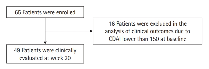
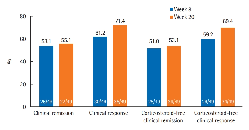
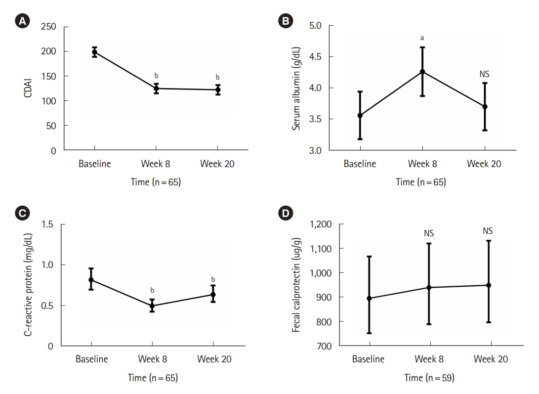
Among 49 patients who had a CDAI of 150 or higher at baseline (Fig. 1), clinical remission and clinical response at week 8 were achieved in 26 (53.1%) and 30 (61.2%) patients, respectively. At week 20, 27 (55.1%) and 35 (71.4%) patients achieved clinical remission and clinical response, respectively (Fig. 2). Corticosteroid-free clinical remission/response were achieved in 25 (51.0%)/26 (53.1%) patients at week 8 and 29 (59.2%)/34 (69.4%) patients at week 20, respectively (Fig. 2). Among 49 patients, the median CDAI score at baseline was 238.0 (IQR, 214.8-257.0), and the median CDAI scores at week 8 and week 20 were 142.4 (IQR, 90.4-193.1) and 129.5 (IQR, 90.65- 190.11), respectively (both P<0.001) (Fig. 3A). Out of 49 patients, 44 (89.8%) were biologic-exposed, and 5 (10.2%) were naïve to biologics. Among 44 biologic-exposed patients, clinical remission/clinical response rates were 47.7%/56.8% at week 8 and 50.0%/68.2% at week 20, respectively. Corticosteroid-free clinical remission/response rates among those 44 patients were 45.5%/54.5% at week 8 and 47.7%/65.9% at week 20, respectively. Among 5 biologic-naïve patients, clinical remission and response rates at weeks 8 and 20 were all 100%. Corticosteroid-free clinical remission/response rates for those 5 patients were also all 100% at weeks 8 and 20.

3. Changes in Biochemical Markers

Relative to baseline, the mean serum albumin level showed a significantly higher level (P<0.05) at week 8 but not at week 20 (Fig. 3B). In the case of CRP, the mean levels at week 8 and week 20 were both significantly lower (P<0.001) compared with baseline (Fig. 3C). In contrast, the mean fecal calprotectin level (Fig. 3D) did not show significant changes during induction therapy.

4. Predictors of Clinical Remission and Clinical Response at Week 20

In the multivariable analysis, underweight (BMI < 18.5 kg/m2) (odds ratio [OR], 0.085; 95% confidence interval [CI], 0.014-0.498; P=0.006) and elevated CRP (≥ 0.6 mg/dL) at baseline (OR, 0.133; 95% CI, 0.022-0.823; P=0.030) were inversely associated with clinical remission at week 20 (Table 2). The history of bowel resection surgery (OR, 0.123; 95% CI, 0.019-0.801; P=0.028) was the only factor significantly associated with clinical response at week 20 (Table 3).

5. AEs during UST Induction Therapy

Twenty-seven of 65 patients (41.5%) experienced AEs during UST induction therapy. SAEs occurred in 3 patients (4.6%): 1 patient was hospitalized and underwent CD-related bowel surgery, and 2 patients were hospitalized due to CD exacerbation. Six patients (9.2%) experienced CD exacerbations, and one patient (1.5%) stopped UST due to a poor response. Five patients (7.7%) experienced upper respiratory tract infections. Other AEs, such as arthralgia and fever, occurred in less than 5% of patients. Details of AEs are summarized in Table 4.

DISCUSSION

In this Korean real-world, multicenter, prospective study, the clinical remission and response rates at week 20 after commencing UST induction therapy for patients with active CD were 55.1% and 71.4%, respectively. Underweight and the elevated CRP at baseline were negatively associated with clinical remission at week 20, and the history of bowel resection surgery was negatively associated with the clinical response at week 20. UST induction therapy was generally well tolerated, with only 4.6% of patients experiencing SAEs.
After placebo-controlled randomized clinical trials on the efficacy and safety of UST for patients with CD [9-11], there have been multiple real-world studies on the effectiveness and safety of UST for patients with CD, mostly conducted in the Western world [12-21]. A recent meta-analysis reported clinical remission and response rates at weeks 8 to 16 to be 34% (95% CI, 26%-42%) and 60% (95% CI, 53%-67%), respectively [36]. Most previous studies have used physicians’ assessments [12,21] or Harvy-Bradshaw index evaluations [13-19] and have evaluated clinical outcomes at weeks 8 to 16 after starting induction therapy [12-19,21].
However, because responses to UST induction therapy are scheduled for evaluations at weeks 16 to 20 according to the Korean governmental reimbursement criteria for UST therapy (after intravenous UST at week 0 and subcutaneous UST at week 8), and the first UST maintenance dosing is scheduled for week 20, we assessed the clinical remission and response rates of study patients at week 20. Additionally, because the Korean reimbursement criteria use CDAI-70 (and not Harvy-Bradshaw index) for assessing responses to UST induction therapy, we used CDAI for defining clinical outcomes. In a recent Hungarian multicenter, prospective study, patients were treated with UST using the same dose and schedule as those used in our study, and clinical remission and response rates were evaluated at weeks 16 to 20 using the same CDAI definition used by our study [20]. At week 8, clinical remission and response rates as per CDAI were 57.7% and 78.1%, respectively [20]. At weeks 16 to 20, clinical remission and response rates were 64.7% and 77.9%, respectively [20]. The Hungarian investigators observed slightly higher clinical remission and response rates than we did. This could be explained by the fact that the Hungarian study included all patients with clinical remission at baseline (14.1% of the total study cohort) and by the fact that a substantial proportion of patients were still on corticosteroids at week 8 [20]. In fact, the corticosteroid-free clinical remission rate at week 8 was 43.8%, which was lower than our corresponding finding [20]. Additionally, differences in patient characteristics, including phenotype and previous treatment, might have been associated with different outcomes.
There have been limited studies on the efficacy or effectiveness of UST induction therapy for Asian patients with CD. A Japanese subpopulation analysis of phase 3 induction and maintenance studies showed generally consistent results with those in the overall population [37]. Real-world studies on the effectiveness of UST for Asian patients with CD have previously been limited to Japanese populations [23-25]. In a retrospective, observational, single-center study of 47 patients with CD, clinical remission rates (CDAI < 150) at weeks 8 and 24 were 44.4% (8/18 patients with CDAI ≥ 150 at baseline) and 66.7% (12/18 patients with CDAI ≥ 150 at baseline), respectively [23]. In another study in which 22 patients with moderate-to-severe CD received intravenous UST therapy every 8 weeks, the rates of clinical remission and response rates were 31.8% and 59.1% at week 8 and 45.5% and 68.2% at week 24, respectively [24]. In an interim analysis of post-marketing surveillance data from Japan, among 130 CD patients with CDAI ≥ 150 at baseline, clinical remission (CDAI ≤ 150) and clinical response (decrease in CDAI ≥ 100 points) were observed in 48.5% and 40% of patients, respectively [25]. Our study provides more real-world evidence for the effectiveness of UST treatment for East Asian patients with CD.
In the present study, there was a significant decrease in CDAI during UST induction therapy. Serum albumin was significantly higher at week 8 compared with baseline, but no significant difference was noted at week 20; serum CRP was significantly lower than baseline at both week 8 and week 20; in contrast, fecal calprotectin did not show significant differences from baseline during UST induction therapy. Previous studies have yielded conflicting results regarding changes in serum CRP during UST induction therapy: decreasing CRP in several studies [12,15,17,23,38-42] and no significant change of CRP in others [14,43]. Conflicting results have also been observed for fecal calprotectin, with decreasing fecal calprotectin [15,39,41,42] or no significant change in this marker [14,43]. Further studies with more patients will be required to confirm the biochemical response during UST induction treatment.
In the present study, underweight at baseline and elevated CRP at baseline were inversely associated with clinical remission at week 20. Also, the history of bowel resection surgery was inversely associated with the clinical response at week 20. Both underweight and elevated CRP might reflect CD activity, leading to poor responses to UST [44,45]. In the case of underweight, Liefferinckx et al. [17] also found that a BMI < 18 kg/m2 was negatively associated with clinical remission at 1 year of UST therapy among CD patients with prior exposure to biologics. In contrast, Wong et al. [46] found that underweight (BMI < 18.5 kg/m2) had no impact on clinical efficacy at week 44 in a post hoc analysis of the IM-UNITI trial. These contradictory results have also been observed regarding anti-TNF therapy for CD patients [47]. In line with our results, Dulai et al. [48] reported that high baseline serum albumin, never smoking, the absence of prior exposure to anti-TNF agents, the absence of active fistulizing disease at baseline, and the absence of prior bowel surgery were predictive of clinical remission at week 16 of UST therapy. Other positive predictors of response or remission after UST induction therapy were UST therapy due to adverse effects of biologics [15,19],UST initiation due to secondary failure to biologics [19], as well as primary nonresponse to anti-TNFs [38] and concomitant immunosuppressant therapy [12]. Conversely, reported negative predictors include old age [19], smoking [19], a higher number of previous anti-TNF agents [15], and endoscopic CD severity [15].
We reported AEs and SAEs in 41.5% and 4.6% of study patients, respectively. These rates were consistent with previous observations. In the UNITI trials, AEs and SAEs were observed in 59.5% and in 5.1% of UST-treated patients, respectively [22]. In a subpopulation analysis of the UNITI trial, AEs occurred in 46.4% and SAEs in 3.6% of Japanese UST-treated patients [37]. In a recent systematic review and meta-analysis of observational studies, among UST-treated IBD patients, AEs and SAEs were observed in 16.7% (498/1,977) and 5.6% (89/1,534) of patients [36]. Although reported rates of AEs and SAEs vary by study, probably due to definitions of events and underreporting, our study adds more evidence for the safety profile of UST for Asian IBD patients.
This was the first study investigating the effectiveness and safety of UST induction therapy for Korean patients with CD. The data were prospectively collected from 4 centers using strictly predefined outcome definitions. However, there were several limitations to this study. First, the sample size was small. For this reason, the statistical power for each analysis may be insufficient, and it was particularly difficult to detect significant changes in biochemical marker levels. Second, because patients were enrolled from 4 centers, there might have been institution-specific differences in management strategies. However, because biologics treatment for patients with CD is strictly regulated by a single universal reimbursement criterion by the Health Insurance Review and Assessment Service of Korea, the general management strategy might not have varied substantially between centers. Moreover, some heterogeneity between hospitals might better reflect the real-world situation. Third, we could not present the data on endoscopic outcomes after UST induction therapy. This is because in Korea, the reimbursement criteria for evaluating UST induction therapy are based on CDAI, which does not require endoscopic evaluation. Therefore, endoscopic evaluations are rarely performed after UST induction therapy for patients with CD in the real-world practice of Korea. Finally, the follow-up duration was relatively short because we only analyzed the outcome of UST induction therapy. We plan to analyze the results of maintenance therapy with a longer follow-up duration in a subsequent study.
In conclusion, UST induction therapy was clinically effective for Korean patients with CD. The safety profile during UST induction therapy was acceptable with no new AEs. In future studies, it will be necessary to analyze the long-term effectiveness and safety of UST maintenance therapy for Asian patients with CD.

ADDITIONAL INFORMATION

Funding Source

This work was supported by the National Research Foundation of Korea (NRF) grant funded by the Korean government (MSIT) (NRF-2021R1A2C2095096).

Conflict of Interest

Ye BD has received a research grant from Celltrion and Pfizer Korea; consulting fees from Abbvie Korea, Celltrion, Chong Kun Dang Pharm., CJ Red BIO, Daewoong Pharma., Ferring Korea, Janssen Korea, Kangstem Biotech, Medtronic Korea, Pfizer Korea, Shire Korea, Takeda Korea, IQVIA, Cornerstones Health, and Takeda; speaking fees from Abbvie Korea, Celltrion, Ferring Korea, Janssen Korea, Pfizer Korea, Shire Korea, Takeda Korea, and IQVIA. Yang SK has received a research grant from Janssen Korea. None of the above-mentioned grants are related to this study. Other authors have nothing to declare.

Kim YS is an editorial board member of the journal but was not involved in the peer reviewer selection, evaluation, or decision process of this article. No other potential conflicts of interest relevant to this article were reported.

Data Availability Statement

Not applicable.

Author Contribution

Conceptualization: Ye BD. Project administration: Ye BD. Data collection: Oh K, Hong HS, Ham NS, Park SH, Yang SK, Yoon H, Kim YS, Choi CH, Ye BD. Analysis and interpretation of the data: Oh K, Hong HS, Lee J, Ye BD. Writing - original draft: Oh K, Hong HS. Writing - review and editing: Ye BD. Approval of the final manuscript: Oh K, Hong HS, Ham NS, Lee J, Park SH, Yang SK, Yoon H, Kim YS, Choi CH, Ye BD. Approval of final manuscript: all authors.

Non-Author Contribution

Dr. Joon Seo Lim from the Scientific Publications Team at Asan Medical Center provided editorial assistance in preparing this manuscript.

Fig. 1.
Flowchart of patient enrollment. CDAI, Crohn’s Disease Activity Index.
ir-2021-00173f1.jpg
Fig. 2.
Clinical outcomes at week 8 and week 20 among 49 patients with Crohn’s Disease Activity Index score 150 or over at baseline.
ir-2021-00173f2.jpg
Fig. 3.
Changes in Crohn’s Disease Activity Index (CDAI) and laboratory values over time. CDAI (A), serum albumin level (B), serum C-reactive protein level (C), and fecal calprotectin level (D) are expressed in means±standard error of the means. The levels of C-reactive protein and fecal calprotectin were log-transformed, analyzed, and back-transformed. aP<0.05, bP<0.001. NS, not significant.
ir-2021-00173f3.jpg
Table 1.
Baseline Patient Characteristics
Variable Value (n = 65)
Age (yr) 33.0 (27.0-45.0)
Male sex 48 (73.8)
Disease duration from diagnosis to ustekinumab commencement (mo) 114.0 (85.0-177.0)
First-degree family history of inflammatory bowel disease 2 (3.1)
Smoking status
 Never 46 (70.8)
 Past smoker 12 (18.5)
 Current 6 (9.2)
 Unknown 1 (1.5)
Body mass index (kg/m2) 20.1 (18.3-24.5)
Disease location
 Ileum (L1) 13 (20.0)
 Colon (L2) 2 (3.1)
 Ileocolon (L3) 50 (76.9)
Upper gastrointestinal involvement 13 (20.0)
Disease behavior
 Nonstricturing, nonpenetrating (B1) 14 (21.5)
 Stricturing (B2) 26 (40.0)
 Penetrating (B3) 25 (38.5)
Perianal disease modifier 32 (49.2)
History of bowel resection surgery 36 (55.4)
Surgical history for perianal abscess/fistula 36 (55.4)
Concomitant immunomodulators 40 (61.5)
Concomitant systemic corticosteroids 5 (7.7)
Prior use of biologics
 Biologic-naïve 7 (10.8)
 Anti-TNFs onlya 37 (56.9)
 Anti-TNFs+vedolizumabb 21 (32.3)
CDAI 209.8 (128.8-240.9)
Clinical disease activity
 Remission (CDAI < 150) 16 (24.6)
 Mild (150 ≤ CDAI < 220) 13 (20.0)
 Moderate (220 ≤ CDAI < 450) 36 (55.4)
 Severe (CDAI ≥ 450) 0
White blood cell (/μL) 6,280 (4,599-8,400)
Hemoglobin (g/dL) 12.4 (11.3-13.5)
Serum albumin (g/dL) 3.5 (3.2-3.8)
Serum C-reactive protein (mg/dL) 0.91 (0.45-1.88)
Fecal calprotectin (mg/kg) 1,304 (394-2,011)

Values are presented as median (interquartile range) or number (%).

a Infliximab (including infliximab biosimilar) for 22 patients, adalimumab for 3 patients, and infliximab (including infliximab biosimilar)+adalimumab for 12 patients.

b Infliximab (including infliximab biosimilar)+vedolizumab for 10 patients, adalimumab+vedolizumab for 1 patient, and infliximab (including infliximab biosimilar)+adalimumab+vedolizumab for 10 patients.

TNF, tumor necrosis factor; CDAI, Crohn’s Disease Activity Index.

Table 2.
Univariable and Multivariable Analysis for Predicting Clinical Remission at Week 20 (n=49)
Variable Univariable
Multivariable
OR (95% CI) P-value OR (95% CI) P-value
Age (per 1 yr) 1.036 (0.988-1.086) 0.145
Female sex 1.123 (0.322-3.919) 0.856
Disease duration (per 1 mo) 0.993 (0.984-1.002) 0.144
Smoking status 0.984
 Never smoker Reference
 Past smoker 0.987 (0.225-4.330) 0.986
 Current smoker 1.184 (0.175-8.021) 0.862
Body mass index
 < 18.5 kg/m2 0.104 (0.024-0.451) 0.002a 0.085 (0.014-0.498) 0.006
 ≥ 18.5 kg/m2 Reference
Disease location 0.178
 Ileum (L1) Reference
 Colon (L2) 999.999 (0.000-999.999) > 0.999
 Ileocolon (L3) 0.253 (0.059-1.078) 0.063
Upper GI involvement 0.373 (0.093-1.496) 0.164
Disease behavior 0.088a
 Nonstricturing, nonpenetrating (B1) Reference
 Stricturing (B2) 3.333 (0.663-16.764) 0.144
 Penetrating (B3) 0.795 (0.157-4.022) 0.782
Perianal disease modifier 0.188 (0.055-0.643) 0.008a 0.239 (0.053-1.081) 0.053
Previous use of biologic agents 0.000 (0.000-999.999) 0.999
Concomitant immunomodulators 0.615 (0.195-1.945) 0.408
Concomitant systemic corticosteroids 0.244 (0.023-2.527) 0.237
History of bowel resection surgery 0.317 (0.091-1.106) 0.072a
History of perianal abscess/fistula surgery 0.275 (0.081-0.930) 0.038a
CDAI at baseline
 < 220 Reference
 ≥ 220 2.514 (0.683-9.252) 0.165
Serum albumin at baseline (per 1 g/dL) 8.829 (1.883-41.391) 0.006a
Elevated CRP (≥ 0.6 mg/dL) at baseline 0.222 (0.059-0.839) 0.026a 0.133 (0.022-0.823) 0.030

a Variables with P<0.1 in univariable analysis were included in multivariable analysis.

OR, odds ratio; CI, confidence interval; GI, gastrointestinal; CDAI, Crohn’s Disease Activity Index; CRP, C-reactive protein.

Table 3.
Univariable and Multivariable Analysis for Predicting Clinical Response at Week 20 (n=49)
Variable Univariable
Multivariable
OR (95% CI) P-value OR (95% CI) P-value
Age (per 1 yr) 1.047 (0.987-1.109) 0.125
Female 3.130 (0.600-16.327) 0.176
Disease duration (per 1 mo) 0.996 (0.986-1.005) 0.395
Smoking status 0.523
 Never smoker Reference
 Past smoker 0.450 (0.098-2.057) 0.303
 Current smoker 1.440 (0.142-14.653) 0.758
Body mass index
 < 18.5 kg/m2 0.462 (0.126-1.697) 0.244
 ≥ 18.5 kg/m2 Reference
Disease location > 0.999
 Ileum (L1) Reference
 Colon (L2) 999.999 (0.000-999.999) > 0.999
 Ileocolon (L3) < 0.001 (0.000-999.999) 0.999
Upper GI involvement 0.625 (0.150-2.599) 0.518
Disease behavior 0.049a
Nonstricturing, nonpenetrating (B1) Reference
Stricturing (B2) 1.810 (0.248-13.211) 0.559
Penetrating (B3) 0.286 (0.046-1.769) 0.178
Perianal disease modifier 0.182 (0.043-0.771) 0.021a
Previous use of biologic agents 0.000 (0.000-999.999) 0.999
Concomitant immunomodulators 0.424 (0.111-1.610) 0.207
Concomitant systemic corticosteroids 0.108 (0.010-1.146) 0.065a
History of bowel resection surgery 0.198 (0.038-1.018) 0.053a 0.123 (0.019-0.801) 0.028
History of perianal abscess/fistula surgery 0.242 (0.057-1.027) 0.054a
CDAI at baseline
 < 220 Reference 5.124 (0.979-26.836) 0.053
 ≥ 220 8.000 (1.933-33.101) 0.004a
Serum albumin at baseline (per 1 g/dL) 2.937 (0.817-10.563) 0.099a
Elevated CRP (≥ 0.6 mg/dL) at baseline 0.211 (0.041-1.091) 0.064a 0.212 (0.033-1.380) 0.105

a Variables with P<0.1 in univariable analysis were included in multivariable analysis.

OR, odds ratio; CI, confidence interval; GI, gastrointestinal; CDAI, Crohn’s Disease Activity Index; CRP, C-reactive protein.

Table 4.
Adverse Events during Ustekinumab Induction Therapy (n=65)
Adverse events No. (%)
Any adverse events 27 (41.5)
Serious adverse events 3 (4.6)
 IBD-related hospitalization 3 (4.6)
 IBD-related surgery 1 (1.5)
 Death 0
Crohn’s disease exacerbation 6 (9.2)
Upper respiratory tract infection 5 (7.7)
Alopecia 3 (4.6)
Arthralgia 3 (4.6)
Hematochezia 3 (4.6)
Myalgia 2 (3.1)
Nausea and/or vomiting 2 (3.1)
Diarrhea 2 (3.1)
Fever 2 (3.1)
Headache 2 (3.1)
Pruritus 2 (3.1)
Discontinuation of ustekinumab 1 (1.5)
Dizziness 1 (1.5)
Insomnia 1 (1.5)
Paresthesia 1 (1.5)

IBD, inflammatory bowel disease.

REFERENCES

1. Ben-Horin S, Chowers Y. Review article: loss of response to antiTNF treatments in Crohn’s disease. Aliment Pharmacol Ther 2011;33:987-995.
crossref pmid
2. Barber GE, Yajnik V, Khalili H, et al. Genetic markers predict primary non-response and durable response to anti-TNF biologic therapies in Crohn’s disease. Am J Gastroenterol 2016;111:1816-1822.
crossref pmid pmc pdf
3. Gisbert JP, Marín AC, McNicholl AG, Chaparro M. Systematic review with meta-analysis: the efficacy of a second anti-TNF in patients with inflammatory bowel disease whose previous anti-TNF treatment has failed. Aliment Pharmacol Ther 2015;41:613-623.
crossref pmid
4. Gisbert JP, Chaparro M. Use of a third anti-TNF after failure of two previous anti-TNFs in patients with inflammatory bowel disease: is it worth it? Scand J Gastroenterol 2015;50:379-386.
crossref pmid
5. Cohen A, Ahmed N, Sant’Anna A. Ustekinumab for the treatment of refractory pediatric Crohn’s disease: a single-center experience. Intest Res 2021;19:217-224.
crossref pmid pmc pdf
6. Benson JM, Peritt D, Scallon BJ, et al. Discovery and mechanism of ustekinumab: a human monoclonal antibody targeting interleukin-12 and interleukin-23 for treatment of immune-mediated disorders. MAbs 2011;3:535-545.
crossref pmid pmc
7. Peluso I, Pallone F, Monteleone G. Interleukin-12 and Th1 immune response in Crohn’s disease: pathogenetic relevance and therapeutic implication. World J Gastroenterol 2006;12:5606-5610.
pmid pmc
8. Neurath MF. IL-23: a master regulator in Crohn disease. Nat Med 2007;13:26-28.
crossref pmid pdf
9. Sandborn WJ, Feagan BG, Fedorak RN, et al. A randomized trial of ustekinumab, a human interleukin-12/23 monoclonal antibody, in patients with moderate-to-severe Crohn’s disease. Gastroenterology 2008;135:1130-1141.
crossref pmid
10. Sandborn WJ, Gasink C, Gao LL, et al. Ustekinumab induction and maintenance therapy in refractory Crohn’s disease. N Engl J Med 2012;367:1519-1528.
crossref pmid
11. Feagan BG, Sandborn WJ, Gasink C, et al. Ustekinumab as induction and maintenance therapy for Crohn’s disease. N Engl J Med 2016;375:1946-1960.
crossref pmid
12. Wils P, Bouhnik Y, Michetti P, et al. Subcutaneous ustekinumab provides clinical benefit for two-thirds of patients with Crohn’s disease refractory to anti-tumor necrosis factor agents. Clin Gastroenterol Hepatol 2016;14:242-250.
crossref pmid
13. Khorrami S, Ginard D, Marín-Jiménez I, et al. Ustekinumab for the treatment of refractory Crohn’s disease: the Spanish experience in a large multicentre open-label cohort. Inflamm Bowel Dis 2016;22:1662-1669.
pmid
14. Eberl A, Hallinen T, Af Björkesten CG, et al. Ustekinumab for Crohn’s disease: a nationwide real-life cohort study from Finland (FINUSTE). Scand J Gastroenterol 2019;54:718-725.
crossref pmid pdf
15. Iborra M, Beltrán B, Fernández-Clotet A, et al. Real-world short-term effectiveness of ustekinumab in 305 patients with Crohn’s disease: results from the ENEIDA registry. Aliment Pharmacol Ther 2019;50:278-288.
pmid
16. Kubesch A, Rueter L, Farrag K, et al. Short and long-term effectiveness of ustekinumab in patients with Crohn’s disease: realworld data from a German IBD cohort. J Clin Med 2019;8:2140.
crossref pmid pmc
17. Liefferinckx C, Verstockt B, Gils A, et al. Long-term clinical effectiveness of ustekinumab in patients with Crohn’s disease who failed biologic therapies: a national cohort study. J Crohns Colitis 2019;13:1401-1409.
crossref pmid pdf
18. Biemans VB, van der Meulen-de Jong AE, van der Woude CJ, et al. Ustekinumab for Crohn’s disease: results of the ICC Registry, a nationwide prospective observational cohort study. J Crohns Colitis 2020;14:33-45.
crossref pmid pmc pdf
19. Casas Deza D, García López S, Lafuente Blasco M, et al. Efficacy and safety of ustekinumab in real clinical practice: retrospective multicentre study. ARAINF cohort. Gastroenterol Hepatol 2020;43:126-132.
crossref pmid
20. Gonczi L, Szanto K, Farkas K, et al. Clinical efficacy, drug sustainability and serum drug levels in Crohn’s disease patients treated with ustekinumab: a prospective, multicenter cohort from Hungary. Dig Liver Dis 2022;54:207-213.
crossref pmid
21. Plevris N, Fulforth J, Siakavellas S, et al. Real-world effectiveness and safety of ustekinumab for the treatment of Crohn’s disease: the Scottish ustekinumab cohort. J Gastroenterol Hepatol 2021;36:2067-2075.
crossref pmid pdf
22. Sandborn WJ, Feagan BG, Danese S, et al. Safety of ustekinumab in inflammatory bowel disease: pooled safety analysis of results from phase 2/3 studies. Inflamm Bowel Dis 2021;27:994-1007.
crossref pmid pmc pdf
23. Miyazaki T, Watanabe K, Kojima K, et al. Efficacies and related issues of ustekinumab in Japanese patients with Crohn’s disease: a preliminary study. Digestion 2020;101:53-59.
crossref pmid pdf
24. Murate K, Maeda K, Nakamura M, et al. Endoscopic activity and serum TNF-α level at baseline are associated with clinical response to ustekinumab in Crohn’s disease patients. Inflamm Bowel Dis 2020;26:1669-1681.
crossref pmid pdf
25. Yokoyama S, Asano T, Nagano K, et al. Safety and effectiveness of ustekinumab in Crohn’s disease: interim results of post-marketing surveillance in Japan. J Gastroenterol Hepatol 2021;36:3069-3076.
crossref pmid pmc pdf
26. Park SH, Kim YJ, Rhee KH, et al. A 30-year trend analysis in the epidemiology of inflammatory bowel disease in the Songpa-Kangdong district of Seoul, Korea in 1986-2015. J Crohns Colitis 2019;13:1410-1417.
crossref pmid pdf
27. Kaibullayeva J, Ualiyeva A, Oshibayeva A, Dushpanova A, Marshall JK. Prevalence and patient awareness of inflammatory bowel disease in Kazakhstan: a cross-sectional study. Intest Res 2020;18:430-437.
crossref pmid pmc pdf
28. Sood A, Kaur K, Mahajan R, et al. Colitis and Crohn’s Foundation (India): a first nationwide inflammatory bowel disease registry. Intest Res 2021;19:206-216.
crossref pmid pmc pdf
29. Sood A, Kaur K, Singh A, et al. Trends of inflammatory bowel disease at a tertiary care center in northern India. Intest Res 2021;19:282-290.
crossref pmid pmc pdf
30. Liu JZ, van Sommeren S, Huang H, et al. Association analyses identify 38 susceptibility loci for inflammatory bowel disease and highlight shared genetic risk across populations. Nat Genet 2015;47:979-986.
crossref pmid pmc pdf
31. Yang SK. How does the epidemiology of inflammatory bowel disease differ between East and West? A Korean perspective. Inflamm Intest Dis 2017;2:95-101.
crossref pmid pmc pdf
32. Jung S, Ye BD, Lee HS, et al. Identification of three novel susceptibility loci for inflammatory bowel disease in Koreans in an extended genome-wide association study. J Crohns Colitis 2021;15:1898-1907.
crossref pmid pdf
33. Yang SK, Park M, Lim J, et al. Contribution of IL23R but not ATG16L1 to Crohn’s disease susceptibility in Koreans. Inflamm Bowel Dis 2009;15:1385-1390.
crossref pmid
34. Zhang J, Chen J, Gu J, Guo H, Chen W. Association of IL23R and ATG16L1 with susceptibility of Crohn’s disease in Chinese population. Scand J Gastroenterol 2014;49:1201-1206.
crossref pmid
35. Silverberg MS, Satsangi J, Ahmad T, et al. Toward an integrated clinical, molecular and serological classification of inflammatory bowel disease: report of a Working Party of the 2005 Montreal World Congress of Gastroenterology. Can J Gastroenterol 2005;19 Suppl A:5A-36A.
crossref pmid pdf
36. Honap S, Meade S, Ibraheim H, Irving PM, Jones MP, Samaan MA. Effectiveness and safety of ustekinumab in inflammatory bowel disease: a systematic review and meta-analysis. Dig Dis Sci 2022;67:1018-1035.
crossref pmid pdf
37. Hibi T, Imai Y, Murata Y, Matsushima N, Zheng R, Gasink C. Efficacy and safety of ustekinumab in Japanese patients with moderately to severely active Crohn’s disease: a subpopulation analysis of phase 3 induction and maintenance studies. Intest Res 2017;15:475-486.
crossref pmid pmc pdf
38. Greenup AJ, Rosenfeld G, Bressler B. Ustekinumab use in Crohn’s disease: a Canadian tertiary care centre experience. Scand J Gastroenterol 2017;52:1354-1359.
crossref pmid
39. Verstockt B, Dreesen E, Noman M, et al. Ustekinumab exposure-outcome analysis in Crohn’s disease only in part explains limited endoscopic remission rates. J Crohns Colitis 2019;13:864-872.
crossref pmid
40. Lorenzo González L, Valdés Delgado T, Vázquez Morón JM, et al. Ustekinumab in Crohn’s disease: real-world outcomes and predictors of response. Rev Esp Enferm Dig 2022;114:272-279.
pmid
41. Monin L, Dubois S, Reenaers C, et al. Ustekinumab in bio-naïve and bio-failure Crohn’s disease patients: Results from a « real-life » monocentric cohort. Dig Liver Dis 2021;53:72-78.
crossref pmid
42. Forss A, Clements M, Myrelid P, et al. Prospective observational study on Stelara (ustekinumab) assessing effectiveness in Crohn’s disease (PROSE): a 16-week follow-up. Scand J Gastroenterol 2021;56:680-686.
crossref pmid
43. Harris RJ, McDonnell M, Young D, et al. Early real-world effectiveness of ustekinumab for Crohn’s disease. Frontline Gastroenterol 2019;11:111-116.
crossref pmid pmc
44. Loly C, Belaiche J, Louis E. Predictors of severe Crohn’s disease. Scand J Gastroenterol 2008;43:948-954.
crossref pmid
45. Agrawal M, Spencer EA, Colombel JF, Ungaro RC. Approach to the management of recently diagnosed inflammatory bowel disease patients: a user’s guide for adult and pediatric gastroenterologists. Gastroenterology 2021;161:47-65.
crossref pmid pmc
46. Wong EC, Marshall JK, Reinisch W, Narula N. Body mass index does not impact clinical efficacy of ustekinumab in Crohn’s disease: a post hoc analysis of the IM-UNITI Trial. Inflamm Bowel Dis 2021;27:848-854.
crossref pmid pdf
47. Gisbert JP, Chaparro M. Predictors of primary response to biologic treatment [anti-TNF, vedolizumab, and ustekinumab] in patients with inflammatory bowel disease: from basic science to clinical practice. J Crohns Colitis 2020;14:694-709.
crossref pmid pdf
48. Dulai P, Guizzetti L, Ma T, et al. Clinical prediction model and decision support tool for ustekinumab in Crohn’s disease. Am J Gastroenterol 2019;114:S373.
crossref
TOOLS
Share :
Facebook Twitter Linked In Google+ Line it
METRICS Graph View
  • 1 Web of Science
  • 1 Crossref
  • 1 Scopus
  • 3,296 View
  • 415 Download
We recommend


ABOUT
ARTICLE & TOPICS
Article Category

Browse all articles >

TOPICS

Browse all articles >

BROWSE ARTICLES
EDITORIAL POLICY
AUTHOR INFORMATION
Editorial Office
Room 310, Lotte Gold Rose II, 31 Seolleung-ro 86-gil, Gangnam-gu, Seoul 06193, Korea
Tel: +82-2-957-6145    Fax: +82-2-957-6146    E-mail: thekasid@irjournal.org                

Copyright © 2024 by Korean Association for the Study of Intestinal Diseases.

Developed in M2PI

Close layer
prev next