INTRODUCTION
Increasing disease burden of IBD in tuberculosis (TB) endemic areas of the globe [1,2], and the similarities between Crohn’s disease (CD) and intestinal tuberculosis (ITB) have continued to perplex the clinicians in deciding the optimal therapeutic strategy for a patient with ulcero-constrictive intestinal disease. Even with the range of available clinical [3], endoscopic [4], histological [5], radiological [6,7], and serological [8] diagnostic modalities, one of the most common approaches remains the administration of empiric antitubercular therapy (ATT), in lieu of possible dissemination of TB with CD specific therapy [9]. However, this approach is not without complications including the hepatotoxic side-effects of ATT, delay in diagnosis of CD, and as reported recently by our group [10], possible worsening of long-term disease course of CD. Hence, in line with these observations, a noninvasive test is need of an hour in order to timely differentiate and start the patient on appropriate CD/ITB specific therapy.
Forkhead box P3 (FOXP3) expressing CD4+CD25+ T-regulatory cells (Treg), the key regulators of mucosal immune response, display a divergent phenotype in chronic inflammatory and infectious disorders with a decreased frequency in the peripheral blood of CD (inflammatory disorder), and an elevated Treg induction in that of pulmonary TB (infectious disorder) patients [11,12]. In our previous study, we have reported that this immunological discrepancy in Treg frequency could be utilized as a blood-based biomarker with a cutoff value of >32.5% to differentiate ITB from CD with a high diagnostic accuracy [13]. The present study was planned to validate the results prospectively in a larger cohort of treatment naïve patients with CD and ITB.
METHODS
1. Patients
Patients with a confirmed diagnosis of ITB, CD, and those with CD/ITB dilemma (indeterminate cases) under follow-up were recruited prospectively from October 2016 to May 2017 at IBD Clinic, All India Institute of Medical Sciences (AIIMS), New Delhi, India. Patients less than 18 years of age, past-history of ATT, with co-existing infection (pulmonary, urinary), HIV seropositivity, pregnant or lactating females, associated malignancy, and those not ready for long-term follow-up, were excluded. The AIIMS Institutional Ethics Committee approved the experimental protocols (approval No. IECPG/484/29.8.16). Written informed consent was obtained from patient population prior to their recruitment.
2. Definitions
1) Crohn’s Disease
The patients were diagnosed as CD based on a combination of clinical, endoscopic and histological features as per the European Crohn’s and Colitis Organisation guidelines [14].
2) Intestinal Tuberculosis
The diagnosis of ITB was made if any of the following criteria was present (1) demonstration of caseating granulomas on biopsy, (2) presence of acid-fast bacillus on the smear examination/culture, (3) demonstration of TB at extrapulmonary site or (4) demonstration of necrotic lymph nodes on computed tomography (CT) chest or abdomen [15].
3) Indeterminate Cases
A therapeutic trial with ATT was given in patients who did not fulfill the above definitions. A patient was categorized as having ITB if patient had clinical and endoscopic/radiologic response to ATT (Paustian’s criteria with Logan’s modification) with a minimum follow-up of 12 months after completion of ATT and diagnosis of CD was made if the patient showed no response, worsened or worsened after initial improvement with ATT trial and subsequently showed a clinical and/or endoscopic response to oral steroids/CD specific therapy [16,17].
3. Sample Collection
Ten milliliters of peripheral blood was collected from treatment naïve CD and ITB patients, and also from indeterminate group (prior to ATT) in heparinized vacutainers for peripheral blood mononuclear cells isolation.
1) Immunophenotyping by Flowcytometry
Ficoll density gradient centrifugation was used for isolation of peripheral blood mononuclear cells and subsequently stained for surface markers with anti-CD4 phycoerythrin, anti-CD25 allophycocyanin and anti-CD127 phycoerythrin Cy7 for 30 minutes at room temperature (BD Biosciences, San Diego, CA, USA). Thereafter, the cells were fixed prior to permeabilization with 1× permeabilization buffer (eBioscience, San Diego, CA, USA), followed by intracellular staining with antiFOXP3 antibodies (eBioscience) for 30 minutes at 4°C. Data were acquired on CyAn (Beckman Coulter, Fullerton, CA, USA) and analyzed using SUMMIT V4.3 software (Beckman Coulter). Results are represented as mean percentage of FOXP3+ cells gated on CD4+CD25+CD127- cells.
4. Statistical Analysis
Categorical variables were expressed as percentage and continuous variables were expressed as mean±standard deviation or median (interquartile range, IQR). Chi-square test was used to compare categorical variables and Student t-test and Mann Whitney U test was used for parametric and nonparametric analysis respectively. Receiver operating characteristics (ROC) curve was constructed to determine area under curve for FOXP3+ cells in peripheral blood. Statistical analysis was done using IBM SPSS statistics 19.0 version (IBM Corp., Armonk, NY, USA) and a P-value <0.05 was considered as statistically significant.
RESULTS
A total of 70 treatment naïve patients with ITB/CD were evaluated between October 2016 till May 2017. Of these 70 patients, 47 were diagnosed with ITB and 23 were diagnosed with CD.
Of 47 patients with ITB, 24 patients (51.1%) satisfied the diagnostic criterion for ITB (11 necrotic lymph nodes on CT abdomen, 9 evidence of active TB on CT chest, 2 both necrotic lymph nodes and active TB on CT chest, 2 both caseating granuloma and active TB on CT chest), and 23 patients were diagnosed after clinical and endoscopic response to ATT. Of 23 patients with CD, 13 were confirmed CD and were treated upfront and 10 were diagnosed with CD after nonresponse to a therapeutic ATT trial and response to CD specific therapy. Hence, 33 patients were indeterminate and were started on therapeutic ATT trial. Of the patients who received ATT, 3 developed adverse events in the form of drug induced hepatoxicity.
1. Baseline Demographic, Clinical, Endoscopic, and Radiologic Characteristics
The mean age at diagnosis and gender distribution was comparable between CD and ITB patients (Table 1), whereas the median disease duration in patients with CD was longer than ITB (36 months, IQR 10-90 months vs. 18 months, IQR 7-36 months; P=0.04). Most common disease location was ileal/ileocaecal in both the groups (Table 1). The frequency of diarrhea (39.1% vs. 13.0%, P=0.01) and blood in stool (30.4% vs. 8.7%, P=0.02) was more common in patients with CD, whereas the frequency of abdominal pain (95.7% vs. 78.3%, P=0.02) was more common in patients with ITB (Table 1). On colonoscopy, left colonic involvement (21.7% vs. 4.5%, P=0.03) and longitudinal ulcers (21.7% vs. 4.5%, P=0.03) were more common in CD, while transverse ulcers (61.4% vs. 21.7%, P=0.001) were more common in ITB. The frequency of other endoscopic features were similar between the 2 groups (Table 2). On histology, granulomas were more common in patients with ITB (28.1% vs. 5.3%, P=0.047). On CT enterography, long segment involvement (60.9% vs. 26.8%, P=0.007) was more common in patients with CD, while necrotic lymph nodes were seen only in patients with ITB (29.5% vs. 0%, P=0.006). Evidence of active TB on CT chest was also seen only in patients with ITB (35.1% vs. 0%, P=0.004) (Table 3).
2. Elevated FOXP3+ T Cells in Peripheral Blood of ITB Patients
The frequency of CD4+CD25+CD127-FOXP3+ T cells in peripheral blood was significantly higher in ITB as compared to CD patients (40.9 [IQR, 33-50] vs. 24.9 [IQR, 14.4-29.6], P<0.001). To predict the cutoff frequency of peripheral CD4+CD25+CD127-FOXP3+ cells for differentiating between ITB and CD, ROC curve was drawn which revealed an area under the curve (AUC) of 0.77 (95% confidence interval [CI], 0.65-0.89; P<0.001) (Fig. 1). According to the coordinates of ROC curve, a cutoff value of 31.35% FOXP3 cells in peripheral blood could predict a diagnosis of ITB with a sensitivity of 83%, specificity of 82.6%, positive predictive value of 91% and a negative predictive value of 68%.
3. Accuracy of FOXP3+ Enumeration in Indeterminate Cases
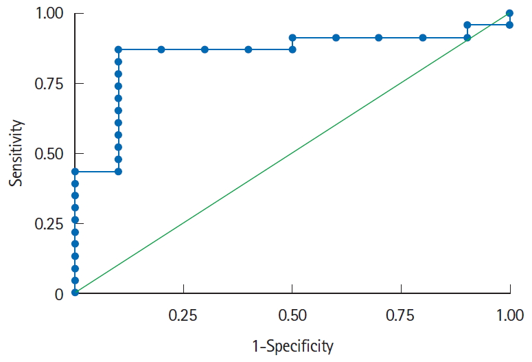
Similar to the entire cohort, CD4+CD25+CD127-FOXP3+ T cells in peripheral blood was significantly higher in patients eventually diagnosed with ITB as compared to those diagnosed with CD (44.3 [IQR, 33.6-53.9] vs. 22.5 [IQR, 14.3-25.8], P=0.005). To predict the cutoff frequency of peripheral CD4+CD25+CD127-FOXP3+ cells for differentiating between ITB and CD, ROC curve was drawn which revealed an AUC of 0.85 (95% CI, 0.68-0.95; P<0.001) (Fig. 2). According to the coordinates of ROC curve, a cutoff value of 32.37% FOXP3 cells in peripheral blood could predict a diagnosis of ITB with a sensitivity of 87%, specificity of 90%, positive predictive value of 95% and a negative predictive value of 75%.
DISCUSSION
Though heterogeneous, in general the literature on FOXP3+ Treg in patients with CD suggests lower frequencies in the peripheral blood and higher in the intestinal biopsy of patients with active CD as compared to controls and patients with inactive disease [18-20]. During disease activity, these regulatory cells are sequestered to the sites of active inflammation, which would account for these observations. On the contrary, infectious disorders such as pulmonary TB have consistently demonstrated an upregulation of these cells in the peripheral blood [11,12], and extending on these observations, we tried to reciprocate these findings in patients with ITB, and compare them with CD. In our previous study, we demonstrated an increased expression of FOXP3 mRNA in the intestinal biopsies of ITB as compared to CD, although there was no difference in the peripheral blood, possibly because of low sample size [21]. Further, in a relatively larger cohort, we showed similar results in the biopsies, along with significantly higher frequency of FOXP3+ Treg cells in the peripheral blood of ITB as compared to CD. Like pulmonary TB, intestinal TB is an infectious disease. Therefore, there is an active expansion of CD4+CD25+ FOXP3+ T cells in ITB patients whose sole role is to balance out the effector responses which is essential to control the ongoing TB infection [22]. However, the role of Treg in CD (where it is generally reduced in the peripheral blood in active CD than controls and inactive CD) which is associated with aberrant immune response is still not clear. A plausible reason to such discrepancy is that during inflammation there is an increase in FOXP3+ T cells in order to balance out the effector responses, however their inability to decrease inflammation suggest that either they have lost their suppressive capability or they have gained the potential to differentiate into Th17 cells. Further, this difference was translated into a simple biomarker with a specificity of 91% to differentiate ITB from CD [13]. However, the sample size was still small with only 16 patients with ITB, and that too with ileocecal disease location were included in this study. In the present study, we not only validated our previous observation in a larger cohort (47 patients with ITB) but also recruited patients with disease location that extended beyond the ileocaecal area. Even with an increased sample size, our present study demonstrates a similar cutoff value of >31.35% with a sensitivity and specificity of 83% and 82.6% respectively (Table 4). Moreover, even in indeterminate cases, FOXP3+ Treg cell enumeration displayed similar diagnostic potential highlighting a strong diagnostic accuracy of FOXP3+ Treg cell in its potential to differentiate ITB from CD.
Differences in other clinical, endoscopic, histologic, and radiologic parameters matched the previous reports with some discrepancies which could be due to differences in patient numbers and characteristics.
Though, multiple studies on various aspects of differentiating ITB from CD have demonstrated reasonable accuracies of different features, one common conclusion that emerges out from these studies is that a single feature cannot differentiate these two diseases, and a combination of features is required to improve the overall diagnostic accuracy in these patients. Moreover, features exclusive to ITB such as caseating granuloma, positive stain for acid-fast bacillus and positive culture on biopsy, necrotic lymph nodes on CT, and evidence of active extra-intestinal TB are limited by their poor sensitivity [23]. Same was exemplified in the present study, with necrotic lymph nodes and active TB on CT chest being seen in only one-third of patients. Various multi-parametric models which have been developed are again limited by their complexity, lack of validation in different populations and infrequent use in the clinics [24,25].
Hence, a simple noninvasive model which is easy to use in the clinics, and incorporates features that can easily be assessed, would improve the diagnostic accuracy of differentiating ITB from CD. In a recent study we combined the CT features (long segment involvement) with visceral fat quantification into a model which had excellent specificity for diagnosis of CD, but was limited by a sensitivity of 50% [26]. He et al. [27] in a recent prospective study from China incorporated age, transverse ulcer, rectum involvement, skipped involvement of the small bowel, target sign, comb sign, and interferon-gamma release assays in a simple nomogram based model with a good diagnostic accuracy for differentiating CD from ITB. Combination of novel diagnostic modalities such as FOXP3+ Treg enumeration, and CT based differentiation with other significant features may enhance the accuracy of individual features and translate into a simple model which can improve the upfront diagnostic differentiation of ITB from CD.
Though conducted prospectively, the study is still limited by relatively less number of patients with CD, which could be attributed to our stringent criteria of including only treatment naïve patients which itself is a challenge in a tertiary care setup. Though there are no population based studies on the epidemiology of ITB or CD from India, CD burden can be estimated indirectly from hospital-based cohort studies, which suggest an indirect estimate of disease burden and its rise over the years in India [28]. India is endemic for TB and abdominal TB is considered as sixth most frequent site of extrapulmonary TB comprising around 5% of all cases of TB [29]. Secondly, we were not able to conduct multivariate analysis or develop a model incorporating multiple parameters including FOXP3+ Treg. However, this is the present focus of our group and we are working on this aspect. We did not check anti-Saccharomyces cerevisiae antibody (ASCA) or interferon-gamma releasing assay (IGRA) levels, as reports from India have shown that ASCA positive serology plays no role in differentiating CD from ITB [30]. While IGRA shows a pooled sensitivity of 80% in differentiating ITB from CD, however it is predictive of latent TB and not active TB [8]. Therefore, by using IGRA, the clinicians cannot rule in or rule out ITB in patients. We have elucidated the shortcomings of both these markers in our previous published review on factors (clinical, pathological, radiological, and immunological) which can differentiate ITB from CD [23].
To conclude, we validate and confirm our previously observed immunological difference between CD and ITB, and propose its use as a simple bedside test which in combination with other features may solve the dilemma of differentiating ITB from CD.